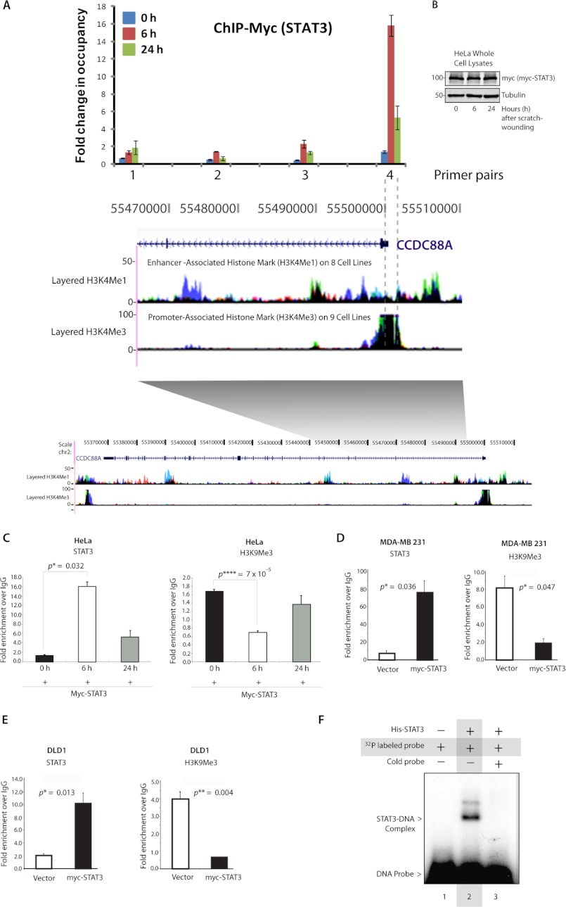
FIGURE 3.
STAT3 binds directly to the GIV promoter. A, top, a schematic representation of STAT3 binding elements on GIV gene is shown. Confluent monolayers of HeLa cells expressing myc-tagged STAT3 were scratch-wounded; harvested at 0, 6, and 24 h postwounding; and analyzed for myc-STAT3 occupancy within a stretch of an ∼3-kb region that includes the GIV promoter and parts of the gene body (indicated by gray shading) by ChIP using anti-myc or control IgGs followed by qPCR using primer pairs that systematically cover the 3-kb region. Shown here are the qPCR results of four such primer pairs, numbered 1–4, demonstrating sites of myc-STAT3 occupancy at different time points during wound healing expressed as -fold enrichment over control IgG. Bottom, a snapshot of the coding region of GIV gene from the UCSC Genome Browser is shown layered below by epigenetic histone marks H3K4me1, which is characteristic of enhancers, and H3K4me3, which is characteristic of promoters. myc-STAT3 binds the GIV gene exclusively at 6 h after wounding and specifically occupies its promoter region (highest enrichment seen with primer pair 4) as determined by its high H3K4me3 and low H3K4me1 associations (demarcated by interrupted gray lines). B, whole cell lysates of HeLa cells used in the above assay were analyzed for expression of myc-STAT3 at 0, 6, and 24 h after scratch wounding by immunoblotting. C–E, increased STAT3 occupancy coincided with a reciprocal loss of the repressive H3K9me3 modification in HeLa (C), MDA-MB 231 (D), and DLD-1 (E) cells. Chromatin immunoprecipitation assays were carried out in HeLa cells (C) after scratch wounding as described earlier and in highly invasive MDA-MB 231 breast cancer (D) and DLD-1 colorectal cancer (E) cell lines overexpressing myc-STAT3 or control vector as indicated using anti-myc (left), anti-H3K9me3 (right), and their respective control IgGs and analyzed for STAT3 and H3K9me3 occupancy by qPCR. F, His-STAT3 binds directly to the GIV promoter as determined by EMSA. Recombinant His-STAT3 formed protein-DNA complexes (shift in lane 2) when incubated with DNA probes harboring the putative STAT3-binding site in the GIV promoter. These complexes are absent in the presence of excess cold probe (lane 3). Error bars represent S.D. *, p < 0.05; **, p < 0.01; ****, p < 0.001.

