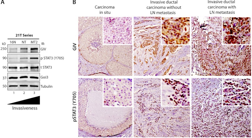
FIGURE 5.
Expression of GIV protein increases during metastatic progression of breast carcinomas. A, whole cell lysates of 21T series of human mammary cells (16N, NT, and MT2) were analyzed for GIV, phospho-Tyr-705 STAT3 (p STAT3), total STAT3 (t STAT3), Gαi3, and tubulin by immunoblotting (IB). B, the abundance of GIV (top) and pSTAT3 (bottom) was analyzed in equal numbers of human breast carcinomas representing three stages during metastatic progression, carcinoma in situ (left), invasive ductal carcinoma without lymph node metastasis (middle), and invasive ductal carcinoma with lymph node metastasis (right). Shown here are representative images of three tumors from separate patients. GIV shows nuclear and cytosolic staining, whereas pSTAT3 is predominantly nuclear. Although carcinomas in situ stained mostly negative for both GIV and pSTAT3, invasive ductal carcinomas frequently stained positive for both (see Tables 1–3). Both the intensity of staining and the percentage of tumor cells that stained positive for GIV and pSTAT3 increased with tumor progression. Insets display a region in the field of the tumor enlarged by digital magnification. Original magnifications, 20×.

