FIGURE 6.
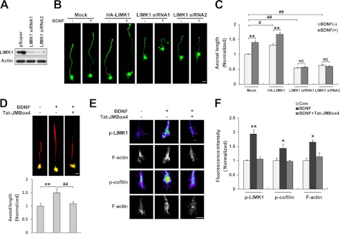
TrkB/LIMK1 interaction is required for BDNF-induced axonal growth. A, LIMK1 siRNA efficiently decreased the expression of endogenous LIMK1. PC12 cells were transiently transfected with LIMK1 siRNA. 72 h after transfection, cells were lysed and immunoblotted with anti-LIMK1 and anti-actin antibodies. B, LIMK1 is required for BDNF-induced axonal elongation. Cultured hippocampal neurons were transfected with HA-LIMK1 or LIMK1 siRNA as described under “Experimental Precedures.” 20 h after BDNF (50 ng/ml) treatment, neurons were fixed and immunostained with rabbit anti-GFP antibodies. Scale bar, 20 μm. C, quantification of axonal length (n = 56–72, **, p < 0.01; Student's t test, #, p < 0.05; ##, p < 0.01; one-way ANOVA). NS, not significant. D, Tat-JMBox4 peptide can block BDNF-induced axonal elongation. Hippocampal neurons were pretreated with Tat-JMBox4 peptide 30 min before BDNF administration. 20 h after BDNF treatment, cells were fixed and immunostained with mouse anti-Tau-1 (red) and chicken anti-MAP2 (green) antibodies. The length of Tau-1-positive neurite was measured as axonal length at DIV3. Representative merged images are shown, and quantification of axonal length is shown below (n = 63–77, **, p < 0.01 versus control group; ##, p < 0.01 versus BDNF-treated group, one-way ANOVA). Scale bar, 20 μm. E, Tat-JMBox4 peptide can block BDNF increased p-LIMK1 and p-cofilin levels and actin polymerization in growth cones. Serum-starved neurons were treated with Tat-JMBox4 peptide (1 μm) and/or BDNF (50 ng/ml for 30 min) followed by fixation and immunostaining. Scale bar, 10 μm. F, quantification of fluorescence intensity of p-LIMK1, p-cofilin, and F-actin (n = 25–38, *, p < 0.05; **, p < 0.01 versus control group; one-way ANOVA).

