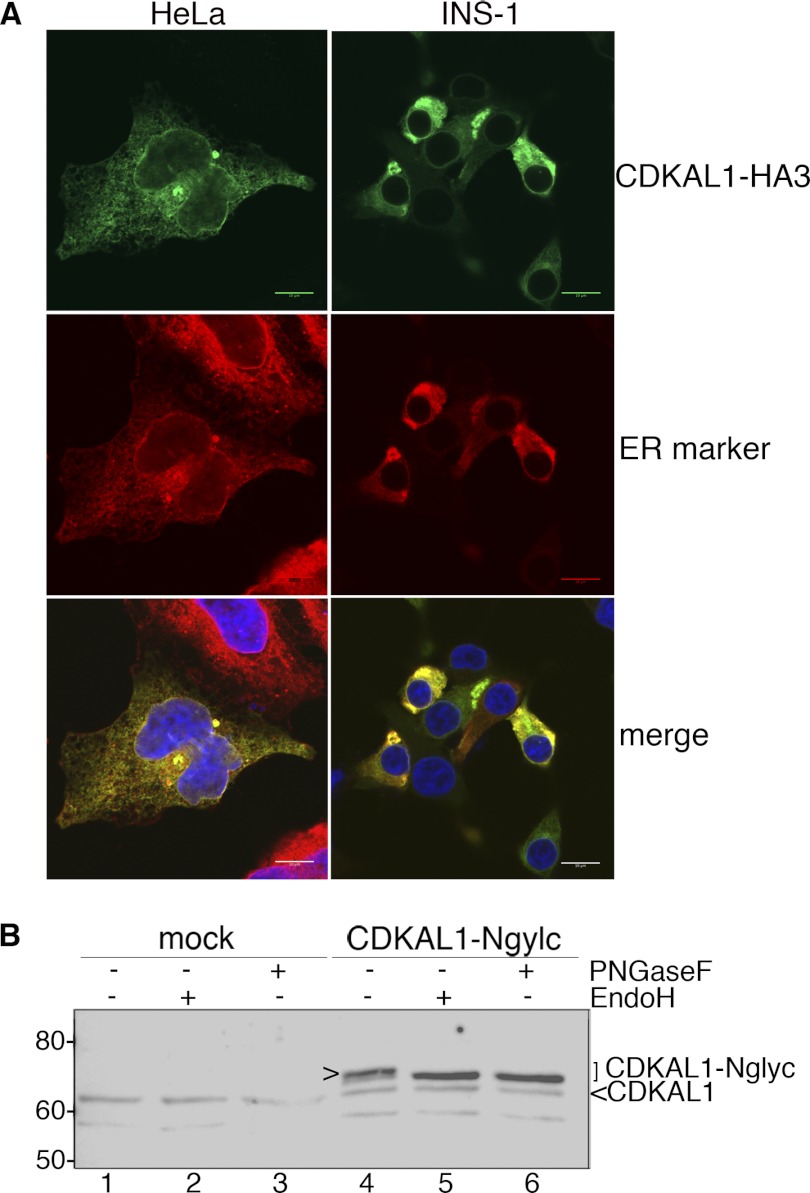
FIGURE 4.
Subcellular localization of CDKAL1 in the ER. A, immunocytochemical detection of CDKAL1-HA3. INS-1 and HeLa cells were transfected with CDKAL1-HA3 and immunostained with the anti-HA antibody (green). In HeLa cells, the ER (red) is visualized by ConA-AlexaFluor594 staining and in INS-1 cells by co-transfection of the fluorescent ER reporter protein mVenus-17 (19). Nuclei are visualized with DAPI (blue). Single confocal sections are shown. Scale bar, 10 μm. B, cell extracts of INS-1 cells transfected with CDKAL1-Nglyc, or the empty vector, were treated with N-endoglycosidases Endo-H and PNGaseF to verify the N-glycosylation of the recombinant protein. Samples were analyzed by SDS-PAGE and immunoblot with anti-CDKAL1 Ab1. Glycosylated CDKAL1-Nglyc is marked with >.

