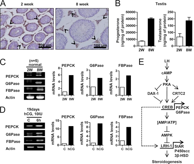
FIGURE 8.
LH increases PEPCK, Glc-6-Pase, and FBPase in mouse testis. A, testes were collected from 2- and 8-week-old C57BL/6J mice for immunohistochemistry assay. The arrowheads show the PEPCK stain in testis. B, testis samples from A was used for RIA. Each point represents the average concentration of progesterone and testosterone from three independent experiments. Data are representative of two independently conducted experiments. C, total RNA was isolated for semiquantitative RT-PCR analysis and real time PCR from 2- and 8-week-old mouse testes. Data represent mean ± S.D. of three individual experiments. D, 19-day-old C57BL/6J mice were injected with hCG (10 IU) for 6 h, and the testes were isolated for semiquantitative RT-PCR analysis and real time PCR. Data represent mean ± S.D. of three individual experiments. E, LH/cAMP/PKA signaling pathway increased expression of PEPCK and Glc-6-Pase via CREB activation, leading to an increased cellular ATP level. Subsequently, AMPK activation decreased in Leydig cells, thereby increasing LRH-1 gene expression, which positively regulates StAR, P450scc, and 3β-HSD steroidogenic enzyme gene expression. Therefore, these effects increase steroidogenesis in mouse testicular Leydig cells.

