FIGURE 1.
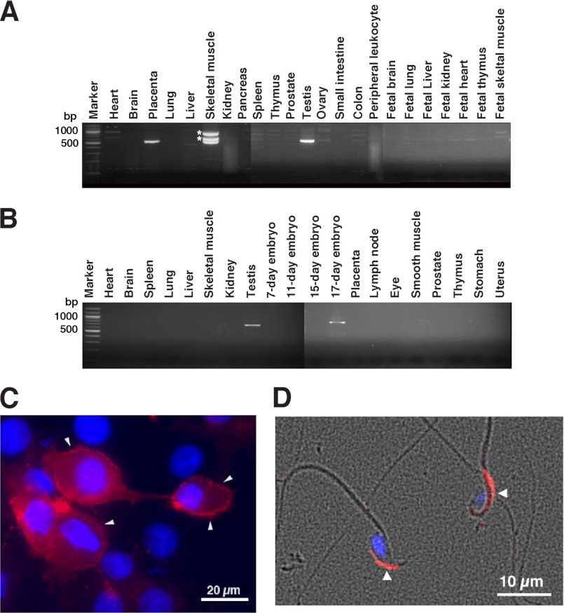
Analysis of the expression pattern of hHYAL4 or mHyal4 mRNA and the cellular localization of mHyal4. The expression pattern of hHYAL4 (A) or mHyal4 (B) mRNA was examined by PCR using cDNA from various tissues. Because three bands were detected in the PCR product using cDNA from human skeletal muscle, they were purified separately from the gel and subjected to the DNA sequence analysis. The upper two bands indicated by asterisks were not derived from the hHYAL4 mRNA but rather nonspecifically amplified PCR bands (data not shown). To examine the cellular localization of mHyal4, COS-7 cells transiently expressing mHyal4 (C) as well as mouse sperm (D) were stained with anti-mHyal4 antibody (red) and DAPI (blue). A merge of the phase contrast and fluorescence images of sperms is depicted in D. The strong staining at the periphery of the COS-7 cells (C, arrowheads) indicates the presence of mHyal4 at the cell surface. The mHyal4 protein was observed in the anterior head of sperm (D, arrowheads).

