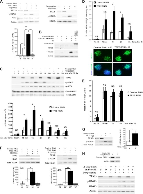
FIGURE 1.
Increased ionizing radiation-dependent phosphorylation of H2AX at DNA double strand breaks upon depletion of TPX2. A, transient transfection of TPX2 siRNA or doxycycline-induced expression of an exogenous miRNA specific for TPX2 significantly increases phosphorylation of H2AX in HeLa cells 1 h after 10 Gy as indicated by Western blot analysis: control siRNA +IR (100.0 ± 13.2) versus TPX2 siRNA +IR (279.0 ± 17.6), p < 0.001, n = 4; −doxycycline +IR (100.0 ± 20.9) versus +doxycycline +IR (871.5 ± 23.4), p < 0.001, n = 4; group (mean ± S.E.), unpaired t test. B, GFP-TPX2 expression reduces the increased levels of γ-H2AX caused by depletion of TPX2 with an siRNA targeting the 3′-untranslated region of TPX2 mRNA. C, time course analysis of H2AX phosphorylation in HeLa cells depleted of TPX2 by siRNA (n = 3–5): 15 min, control siRNA (78.6 ± 31.7) versus TPX2 siRNA (221.3 ± 25.9), p < 0.05; 1 h, control siRNA (90.4 ± 18.5) versus TPX2 siRNA (354.1 ± 54.8), p < 0.05; 2 h, control siRNA (100.0 ± 0.0) versus TPX2 siRNA (206.9 ± 38.4), p < 0.05; group (mean ± S.E.), unpaired t test. Note the similar ATM activation in irradiated control and TPX2-depleted cells as indicated by the levels of p-ATM (Ser-1981). D, increase in the number of U2OS cells with more than five high intensity γ-H2AX ionizing radiation-induced foci (i.e. intensity two standard deviations above the mean focus intensity) following TPX2 depletion by siRNA, 1 and 2 h after 4 Gy: 1 h, control siRNA (1.7 ± 0.4%) versus TPX2 siRNA (6.7 ± 1.1%), p < 0.05, n = 3; 2 h, control siRNA (1.9 ± 0.3%) versus TPX2 siRNA (5.2 ± 0.9%), p < 0.05, n = 3; group (mean ± S.E.), unpaired t test. The number of cells with these high intensity ionizing radiation-induced foci declines 3 h post-IR. Representative pictures of γ-H2AX ionizing radiation-induced foci in control RNAi- and TPX2 RNAi-treated cells 2 h after 4 Gy are shown. E, TPX2 depletion in U2OS cells by siRNA does not cause a significant change in the mean number of γ-H2AX ionizing radiation-induced foci after 4 Gy (n = 3). NS, non-significant, unpaired t test, S.E. F, increased phosphorylation of H2AX 1 h after 10 Gy in caspase-3-deficient MCF-7 cells depleted of TPX2 for 24 or 48 h by siRNA (n = 3) as detected by Western blots. MCF-7 cells do not undergo ionizing radiation-induced apoptosis associated with DNA fragmentation (54, 55). G, increased phosphorylation of H2AX in TPX2-depleted HeLa cells 2 h after a non-lethal dose of 2 Gy (n = 4). H, increased γ-H2AX levels in TPX2-depleted cells 1 h after ionizing radiation are unaffected by the broad spectrum caspase inhibitor Z-VAD-fmk. Z-VAD-fmk was applied at a known effective dose (51) and inhibited PARP1 cleavage (a marker for apoptosis) in the presence of the DNA-damaging drug camptothecin (top blots). Relative quantifications of γ-H2AX signals from independent experiments are shown in bar charts (A, C, F, and G). n = number of independent experiments; NS, non-significant; *, p < 0.05, **, p < 0.01; ***, p < 0.001. Error bars represent S.E. Bar in D, 10 μm. A.U., arbitrary units; Con, control.

