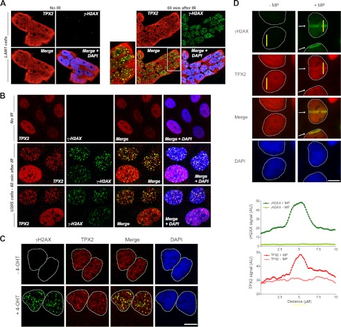
FIGURE 3.
TPX2 localizes to DNA double strand breaks. A and B, TPX2 partially co-localizes with γ-H2AX-positive ionizing radiation-induced foci after 5 Gy in LAN1 (A) and U2OS cells (B), respectively (see also supplemental Fig. 5). TPX2 is found in the nucleus and cytosol in neuroblastoma LAN1 cells and neurons (see text and supplemental Fig. 5 for details). C, TPX2 co-localizes with γ-H2AX at 4-hydroxytamoxifen (4-OHT)/AsiSI-induced DNA double strand breaks. U2OS cells stably expressing AsiSI-estrogen receptor were left untreated or treated with 300 nm 4-hydroxytamoxifen for 4 h and subsequently immunostained for TPX2 and γ-H2AX. D, TPX2 accumulates in DNA double strand break-containing laser tracks (indicated by white arrows) marked by γ-H2AX. U2OS cells were either mock-treated (−MP) or microirradiated with a multiphoton laser (+MP). A representative image shows TPX2 accumulation at 10 min after irradiation. The intensity profiles of γ-H2AX and TPX2 immunofluorescence signals were measured in the yellow bars perpendicular to the laser tracks. Commercially available TPX2 antibody 184 was used in all immunofluorescence images. See supplemental Fig. 5 and Fig. 5 for specificity of TPX2 184 antibody. Bars, 10 μm. AU, arbitrary units.

