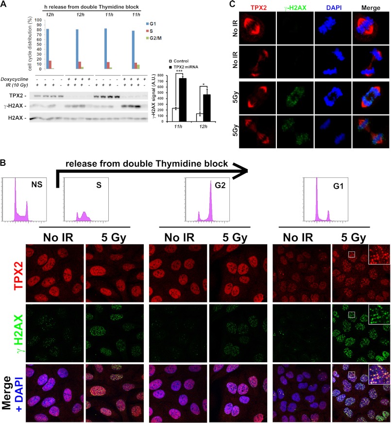
FIGURE 4.
TPX2 regulates the levels of ionizing radiation-dependent γ-H2AX and forms ionizing radiation-induced foci in G1 phase cells. A, enhanced levels of γ-H2AX 1 h after 10 Gy in G1 phase HeLa cells depleted of TPX2 by doxycycline-induced TPX2 miRNA expression. Note that the γ-H2AX augmentation in these TPX2-depleted cells is ionizing radiation-dependent. Cells were synchronized using a double thymidine block, released into fresh medium, and then used at specified time points for ionizing radiation treatment as indicated. Unirradiated cells were used for flow cytometry-based cell cycle profiling (top bar charts). Relative quantifications of γ-H2AX signals from independent experiments are shown in bar charts on the right: 11 h, −doxycycline +IR (229.0 ± 21.0) versus +doxycycline +IR (748.6 ± 39.4), p < 0.001; 12 h, −doxycycline +IR (135.1 ± 34.2) versus +doxycycline +IR (466.1 ± 98.6), p < 0.05; group (mean ± S.E.), unpaired t test; n = 3 (independent experiments); *, p < 0.05; ***, p < 0.001. Error bars represent S.E. Note that 11 h after release from the thymidine block TPX2-depleted cultures contain slightly more G2/M phase cells (8.66%) than control cultures (3.25%). Thus, on the Western blot, a 12 h-11 h loading hierarchy is chosen to facilitate comparison between control 11 h versus +doxycycline 12 h (3.96% G2/M cells). Although the cell cycle profile between these two samples is highly similar, TPX2-depleted cells exhibit approximately twice the levels of γ-H2AX than control cells. B, U2OS cell cultures synchronized with a double thymidine block as in A (see flow cytometry-based cell cycle profiles in top histograms; NS, non-synchronized control) form TPX2 ionizing radiation-induced foci 1 h after irradiation that partially co-localize with γ-H2AX during G1 phase. All images were taken under identical experimental and microscopic conditions. See text for details. C, TPX2 maintains its association with the mitotic spindle in the presence of DNA double strand breaks marked by γ-H2AX. Early and late mitotic figures as identified via DAPI and TPX2 staining with and without DNA damage are shown. Commercially available TPX2 antibody 184 was used in all immunofluorescence images. See supplemental Fig. 5 and Fig. 5 for specificity of TPX2 184 antibody. A.U., arbitrary units.

