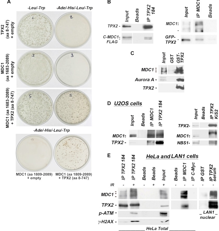
FIGURE 6.
TPX2 associates with MDC1, p-ATM, NBS1, and γ-H2AX. A, Y2H experiment using bait-TPX2 (aa 8–747) and prey-MDC1 (aa 1683–2089 or 1809–2089). Plasmids were co-transformed as indicated. −Leu/−Trp selection agar plates are controls for transformation efficiency. Colonies on −Ade/−His/−Leu/−Trp selection agar plates reveal an interaction between bait-TPX2 and prey-MDC1. Plates were incubated for 6 days. B, an ectopic C-terminal fragment of MDC1 (C-MDC1-FLAG; aa 1807–2089) associates with endogenous TPX2 in HeLa cells as indicated by co-immunoprecipitations with TPX2 antibody 184 from total cell lysate (left panel). Ectopic GFP-TPX2 also co-immunoprecipitates with endogenous MDC1 (right panel). The input lane for the C-MDC1-FLAG (left panel) is from a shorter exposure of the same Western blot. The input lane for the endogenous MDC1 (right panel) is from a stronger exposure of the same Western blot. C, GST-TPX2 pulls down MDC1 and the positive control Aurora A from total HeLa cell lysate. For MDC1, the input is a shorter exposure of the same blot. D, co-immunoprecipitations from U2OS cell lysates with the specified antibodies (see text for details on antibodies). TPX2 was co-immunoprecipitated with MDC1 from these cells using MDC1 antibody (left panel). MDC1 also co-immunoprecipitated with TPX2 antibody 184 (left panel). TPX2 was also found in complex with NBS1 and MDC1 species that migrate slower on SDS-PAGE gels when the co-immunoprecipitations were performed with the TPX2 KiS2 antibody (right panel; see text for further details). E, TPX2 and MDC1 associate in HeLa and LAN1 cells as detected by co-immunoprecipitations with the specified antibodies from total (left and middle panels) or nuclear (right panel) lysates. TPX2 from neuroblastoma LAN1 cells (and primary neurons) migrates as a doublet on gels (see supplemental Fig. 5). TPX2 is also found in complex with p-ATM and γ-H2AX after ionizing radiation treatment. Beads alone or antibodies against c-Myc were used as negative controls as indicated. IP, immunoprecipitation.

