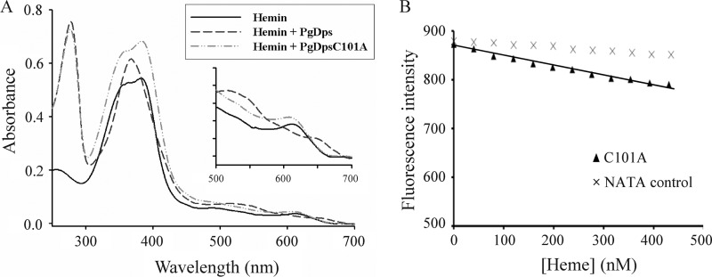
FIGURE 5.
Cysteine-101 of PgDps coordinates the heme iron. A, binding of ferric heme to PgDpsC101A gave similar spectral features to hemin alone, in contrast to the spectral changes induced by native PgDps. B, maximal tryptophan fluorescence quenching of apo-PgDpsC101A at 358 nm, pH 8.0, by sequential titrations of heme. Background quenching was obtained using N-acetyltryptophanamide (NATA) under the same conditions.

