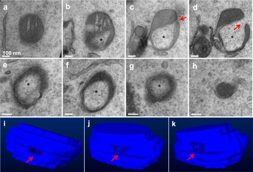
FIGURE 2.
Serial sections of a mitochondrial spheroid. MEFs treated with CCCP for 6 h were fixed for EM examination. When areas enriched for the mitochondrial spheroids were identified, serial sections were performed. Shown in a–h is a series of sections separated by 50 nm in distance. Note the lumen (asterisk) that contains the cytosol and the orifice (arrow) that connects the lumen to the cytoplasm. The serial sections allowed the construction of a three-dimensional model, and representative views of the model are shown in i–k.

