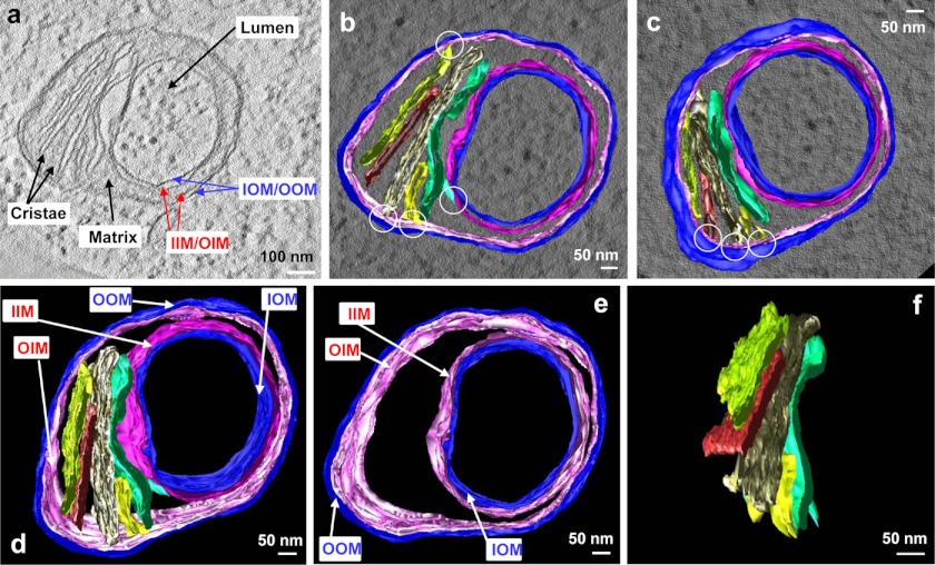
FIGURE 4.
The topological relationship of inner and outer membranes in a mitochondrial spheroid. a, a tomographic slice shows the inner membrane (IM), the outer membrane (OM), and the matrix enclosed by the IM, in which two of the five cristae are also indicated. b–f, the three-dimensional model of the IM (magenta), the OM (blue) and the five cristae (in various colors) were superimposed on a tomographic slice and viewed from the top (b) and bottom (c). Note the connection of the cristae to the IM (white circles). d–f show the model with all of the membrane components (d), the IM and OM only (e), and the cristae only (f). Note the lamellar form of the cristae in f. Images a and b–f were extracted from supplemental Videos 4 and 5, respectively.

