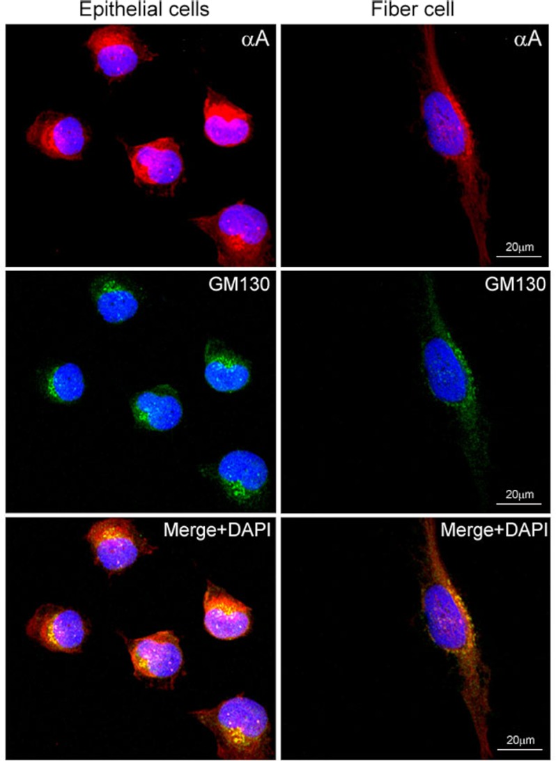
FIGURE 1.
Confocal images of αA localization in primary cultures of rat lens epithelial explants. The P10 rat lens epithelial explants in culture invariably contain nascent (differentiating) fiber cells (14). Perinuclear colocalization of αA (anti-αA, red, top panel) and GM130 (anti-mouse GM130; FITC, green, middle panel) is observed in lens epithelial cells (left column) as well as in the differentiating fiber cell (right column). The αA label (red) is predominantly outside of the Golgi (compare the colocalized yellow granules with the red stain in the bottom panel). Note the granular appearance of the colocalized proteins (bottom panel, yellow, Merge + DAPI) and as yet unrecognized presence of αA (red streaks), prominent in the nucleus in the fiber cell (right bottom panel). Nuclei are stained with DAPI (blue). Scale bar, 20 μm.

