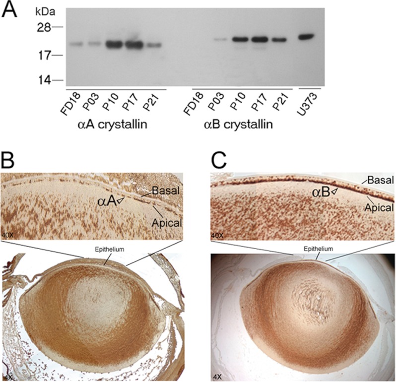
FIGURE 2.
Developmental expression and immunohistochemistry of αA and αB in the native ocular lens. A, immunoblot showing temporal expression pattern of αA and αB during lens development in the rat. Total protein extracts (0.2 μg/lane) from fetal day 18 (FD18) and postnatal days (P3, P10, P17, P21) were analyzed on two immunoblots. αA is expressed early in the FD18 rat lens, when there is no detectable αB. Human glioblastoma cell U373 MG total cell extract (20 μg), which only expresses αB, is shown in the last lane. Protein standards (kDa) are shown on the left. B and C, immunohistochemistry of αA and αB localization, respectively. Immunoperoxidase-diaminobenzidine-stained 4× image of the whole ocular lens is shown in the bottom panels and the central epithelium of this image is magnified (40×) and shown in the upper panels. Note that αA is apical in its location, which suggest its association with the apical Golgi, but there are discontinuities in its staining. The data shown in C confirm previously reported (14) colocalization of αB in the apical Golgi. Note definitive αB staining (C) in the apical epithelium in comparison with anti-αA staining in B (open arrowheads).

