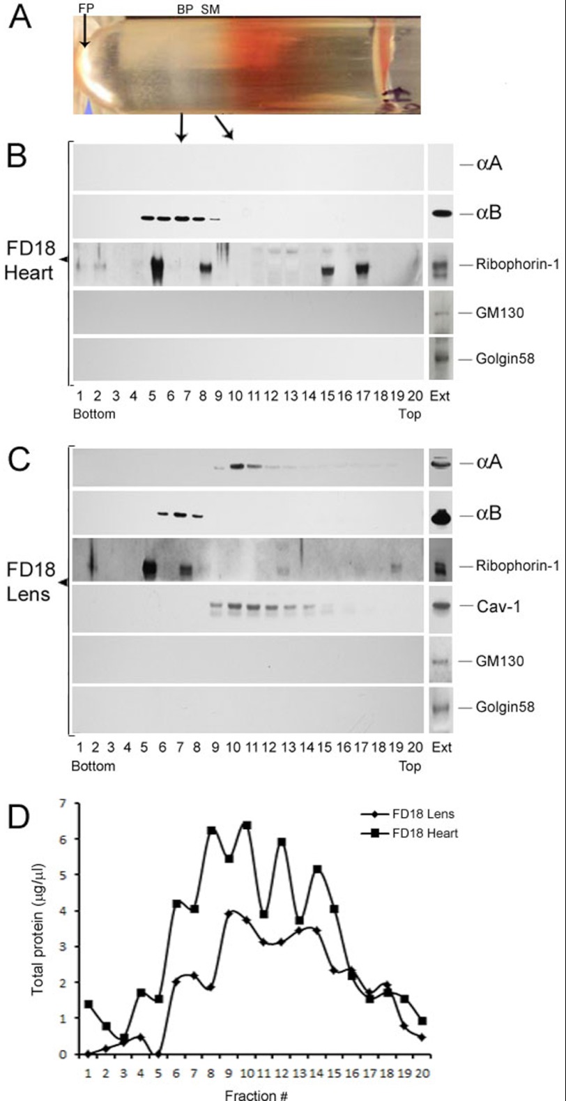
FIGURE 5.
Fractionation of smooth membranes and rough ER in FD18 rat lens and heart. A, picture (shown horizontally) of the fractionated gradient obtained with post mitochondrial supernatant of FD18 heart. The arrows indicate the positions of the bound polysomes (BP, also known as rough ER), the SM and free polysomes (FP). B, immunoblots of the gradient shown in A with anti-αA, anti-αB, anti-Ribophorin-1, anti-GM130, and anti-Golgin 58. Equal volumes (2.5 μl) from each fraction were used for immunoblotting. Note the absence of αA reactivity because there is no αA expressed in the heart (top panel). αB is seen in fractions 5–9 as is Ribophorin-1, which seems to associate with two discrete fractions of the bound polysomes (rough ER). It is also detected in the top half of the gradient in fractions 15 and 17). C, fractionation of FD18 lens post-mitochondrial supernatants. Note that αA (fractions 9–15) fractionates away from αB (fractions 6–8). Caveolin-1 (Cav-1) fractionates with SM in the same location where αA is seen. Note that Ribophorin-1 is mostly seen here with the RER. Transferrin, one more BP/RER marker, could not be detected in this gradient, although it is seen in P10 lens gradients (see Fig. 6). EXT, immune reactivities in aliquots of total cell extracts before fractionation. D, the distribution of total protein in FD18 lens and FD18 heart gradients. Note that the immune reactions seen for αA and αB do not correspond with this distribution pattern.

