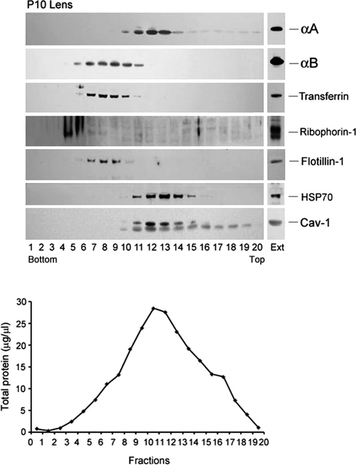
FIGURE 6.

Fractionation of smooth membranes and rough ER from P10 rat lens. A similar gradient as in Fig. 5 was run. αA (top panel) fractionates with smooth membrane fractions 10–15 in which Caveolin-1 and HSP70 are detected (bottom two panels). αB is detected in fractions 5–11 (second panel) distinct from αA, in the rough ER, which is characterized by the presence of the marker Transferrin (third panel), Ribophorin-1 (in heavier polysomes, fourth panel), and Flotillin-1 (fifth panel). There is an overlap of αA and αB patterns in fractions 10 and 11. A light reaction for both αA as well as Caveolin-1 (Cav-1) is seen in the top fractions (15 onwards) in the gradient possibly because of the presence of high concentrations of αA in the ocular lens. EXT, immune reactivities in aliquots of total cell extracts before fractionation. The total protein in each fraction (μg/μl) is plotted in the lower panel. Note that there is no strict correspondence between immune reactions (particularly with αB) and the protein concentration profile.
