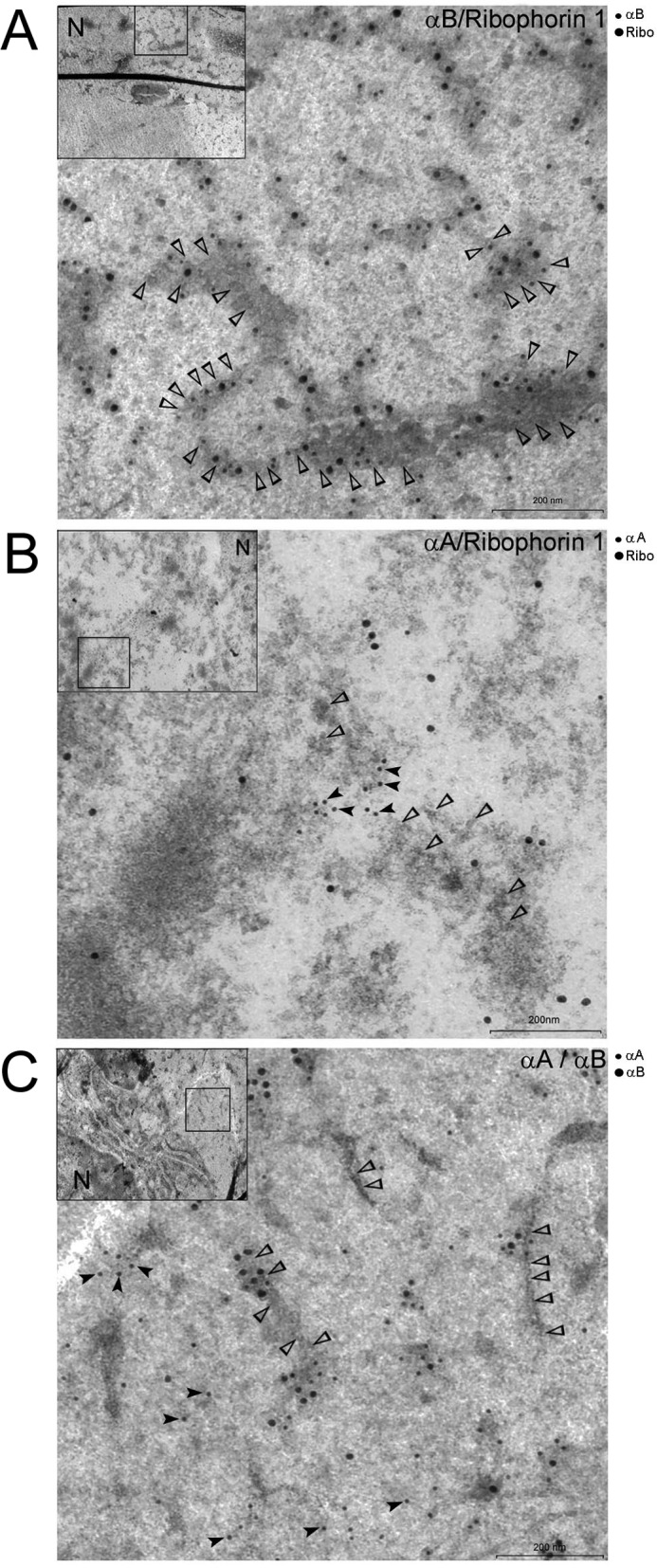
FIGURE 8.

Localization of αB and Ribophorin-1 in native lens fiber cells. Shown above are three representative transmission electron microscopy images of the data obtained with immunogold labeling of αA, αB, and Ribophorin-1 (Ribo) in rat lens fiber cell ultrathin sections. Two antibodies were used for generating each picture as indicated in the top righthand corner of each micrograph. A, localization of αB (12-nm gold) and Ribophorin-1 (18-nm gold). Note that both the proteins are associated with membrane decorated with ribosomes (open arrowheads). B, micrograph showing labeling with anti-αA (12-nm gold, black arrowheads) and anti-Ribophorin-1 (18 nm gold). Note that there are very few 18-nm particles (Ribophorin-1) in the vicinity of 12-nm particles (αA); open arrowheads point to membrane-bound ribosomes. The two proteins do not seem to localize within the same membrane domains. C, micrograph showing labeling with anti-αA (12-nm gold) and anti-αB (18 nm). Open arrowheads point to membrane-bound ribosomes. This micrograph is similar to localization of αA and Ribophorin-1 shown in B. The two proteins do not share the same membrane domains. Low magnification images (insets) are shown in the top left corner of each micrograph. The square box in the inset shows the area magnified. Similar electron micrographs acquired from different regions of fiber cells are presented in supplemental Fig. S2, A–D. Scale bar, 200 nm. N, nucleus. Preimmune serum controls are presented in supplemental Fig. S2D.
