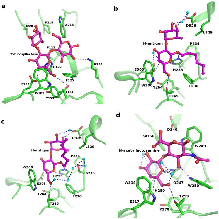
Figure 3. Acceptor binding pocket among GT6 family members showing the conserved residues and interactions.
(a) BoGT6a in complex with FAL; (b) GTA in complex with H-antigen (PDB id: 1LZI); (c) GTB in complex with H-antigen (PDB id: 1LZJ); (d) α3GT in complex with LacNAc (PDB id: 1GX4). Water molecules are shown as cyan spheres.

