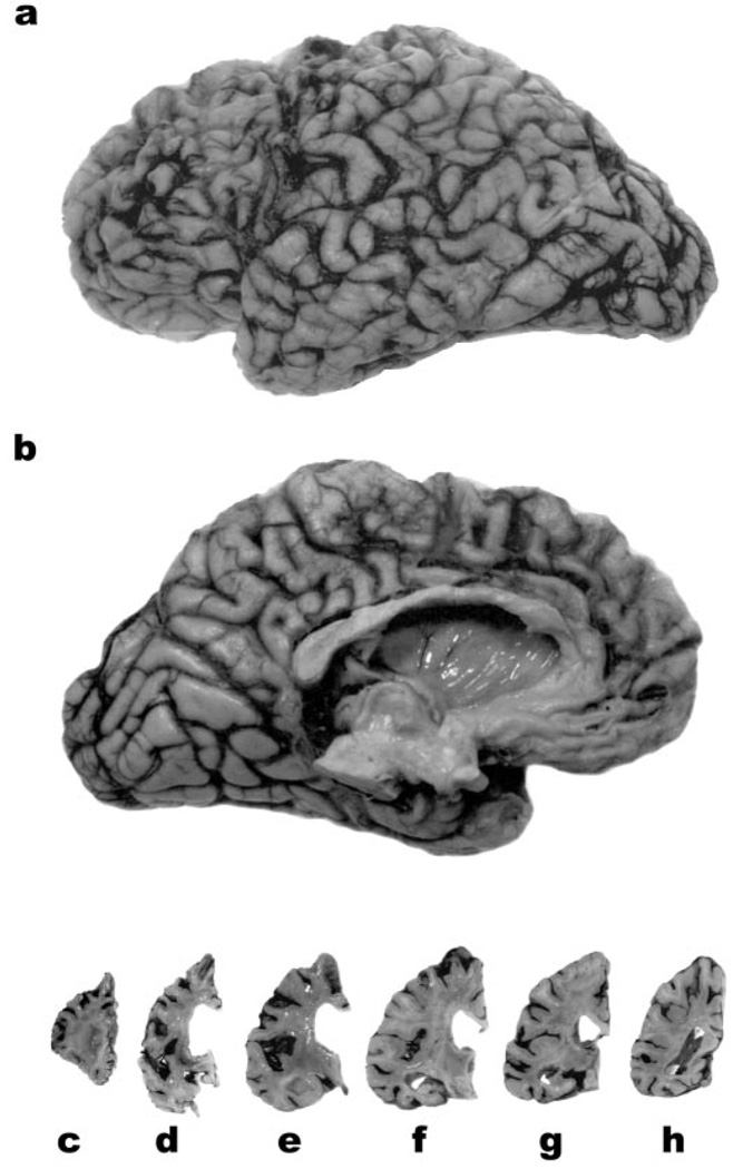
Figure 1.
The lateral (a) and medial (b) aspects of the left hemibrain of 27-year-old woman with neuronal intermediate filament inclusion disease. There is pronounced atrophy of the frontal and anterior temporal lobes. Coronal slices of the left hemisphere (c through h) reveal enlargement of the lateral ventricle, and marked atrophy of the striatum (d). The Sylvian fissure (e) is widened and the amygdala (e) and hippocampus (f, g) are atrophied. The parietal (a, b, h) and occipital (a, b) lobes are relatively well preserved.

