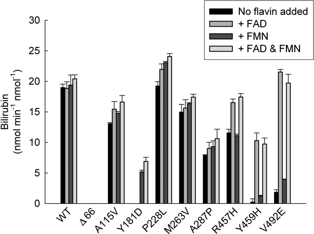
Figure 2. Flavin Activation of CYPOR Variants.
Reconstituted systems were prepared containing each of the CYPOR variants shown ([CYPOR] = [HO-1] = 0.05 µM) and assayed for HO-1 activity in the presence/absence of 20 µM FAD, FMN, or both. Each bar represents the mean ± standard deviation of triplicate assays.

