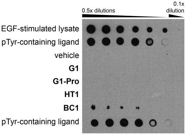
Figure 6.
Anti-pTyr dot blot assay. Peptides were normalized to 750 μM, then serially diluted as shown and spotted onto nitrocellulose. The membranes were blocked with bovine serum albumin, probed with anti-pY antibodies 4G10 (above) or PY20 (see Fig. S4), probed with secondary antibody, and then images were developed using chemoluminescent detection. Controls included EGF-stimulated cell lysate (highest concentration is 1 mg/mL total protein), the dye-labeled, pTyr-containing probe peptide used in the fluorescence polarization assays (pTyr-containing ligand), and vehicle (50% DMSO).

