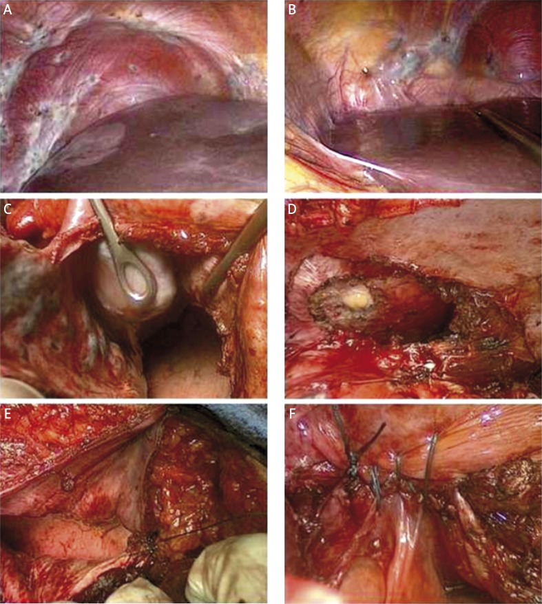
Figure 1.
Right (A) and left (B) diaphragmatic peritoneum widely involved by confluent nodules, from 0.5 cm to 5 cm, infiltrating the diaphragmatic central tendon, close to the upper hepatic veins and on the diaphragmatic pericardial insertion. Right (C) and left (D) wide diaphragmatic resection, with opening of the pleural and pericardial cavity, revealing multiple parietal full-thickness infiltrating nodules involving the parietal pleura, the diaphragmatic central tendon and infiltrating the diaphragmatic side of the pericardium. Right pleural and diaphragmatic suture (E) and left pleural and diaphragmatic suture with pericardial window closure (F) by a single layer (1-0 polypropylene) suture

