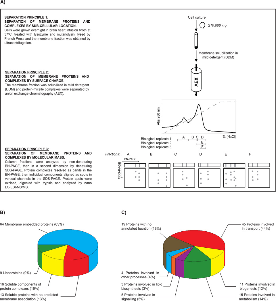
Figure 1. A birds-eye view of the membrane proteome of E. faecalis OG1X.
(A) An overview of the methodology used in this study. A detailed description of all methodology is available in Supplementary Materials and Methods. Identified proteins were classified by (B) cellular location and (C) function.

