
Fig. 5.
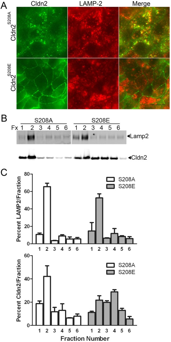
Intracellular S208A mutant of claudin-2 is largely colocalized with LAMP-2, a lysosomal membrane marker. (A) Immunofluorescent co-localisation of claudin-2 mutants expressed in MDCK I cells revealed that claudin-2 S208A (green, top panels) but not S208E (green, bottom panels) colocalized with the lysosomal marker LAMP-2 (red, middle panels). (B) Biochemical enrichment of lysosomes by iodixinal fractionation followed by claudin-2 and LAMP-2 immunoblots demonstrates that claudin-2 S208A concentrates in the same gradient fractions as LAMP-2 (left), while claudin-2 S208E was distributed throughout the gradient. (C) Quantification of the above blot and a separate experiment performed on different claudin-2 S208A and S208E cell clones reveals that most S208A is concentrated in the top two gradient fractions with LAMP-2, while S208E does not show this preferential co-fractionation with the lysosomal marker (mean and range plotted).