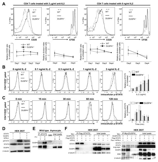
Figure 5.
DUSP4 ablation decreases IL-2 signaling threshold through sustained STAT5 phosphorylation. (A) WT or DUSP4−/− T cells were stimulated and analyzed as in Fig. 4A without additional treatment, or in the presence of neutralizing anti-IL-2 antibody or exogenous IL-2. Representative CD25 (day 1) or CFSE (day 2) flow cytometry results on gated CD4+ cells are shown (top row). Also shown are daily CD25+ percentage and the division index of viable CD4 T cells of treated samples normalized to untreated samples (bottom row, n=7). ** p<0.05. See Supporting Information S5 and S6 for raw data. (B-C) Purified T cells were stimulated as in Fig 4Afor 44 hrs, rested for 4 hrs, and treated with various doses of IL-2 for 15 min (B) or 3 ng/ml IL-2 for different time (C), followed by flow cytometry analyses of CD4, CD25, and p-STAT5 (Y694) (left panels). Mean fluorescence level of p-STAT5 in CD4+CD25+ cells was normalized to that of CD4 to compensate for staining variation, with the results shown as fold-induction to untreated samples (n=4) (right panels). (D) 293T HEK cells were transiently transfected with the indicated vectors. Twenty-four hr later, cells were treated with 10 ng/ml human IFN-β (hIFN-β) for 15 min or left untreated prior to western analyses for STAT5 Y694 phosphorylation (pSTAT5). STAT5, DUSP4, and β-tubulin loading control are also shown. (E) Total lysates from WT thymocytes treated or untreated with 50 ng/ml PMA and 500 ng/ml ionomycin (P+I) for two hours were immuoprecipitated with anti-STAT5 antibody. The precipitated proteins (IP-STAT5), together with aliquots of the original total lysate (total lysate) were analyzed by western analyses for DUSP4 and STAT5. Due to the amount of lysate loaded, DUSP4 was not detectable in lane 3 and 4. MM, molecular-weight marker. (F). 293T HEK cells were transfected and stimulated with 10 ng/ml human interferon-β (hIFN-β) for 1 hour as indicated. Cell lysates were analyzed by western blotting directly (total lysates) or following immunoprecipitation with anti-Flag (left panel) or anti-Myc (right panel) antibody. Perv., 25 μM pervanadate. Representative results from two (panel D and E) or three (panel A–C, and F) independent experiments are shown. p>0.05 unless specified.

