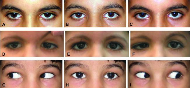
Figure 1. Ocular motility.
Images A, D, and G are right gaze; images B, E, and H are primary gaze; and images C, F, and I are left gaze. Figures A, B, and C show Patient A1 with total horizontal gaze palsy, while Figures D, E, and F show Patient C1 with bilateral DRS type 3, and Figures G, H, and I are Patient C4 with entirely normal ocular motility. All of these patients had full vertical gaze OU. This montage illustrates the spectrum of horizontal gaze with homozygous HOXA1 mutations, from complete horizontal gaze restriction (top row) to DRS type 3 OU (middle row) to full motility (bottom row).

