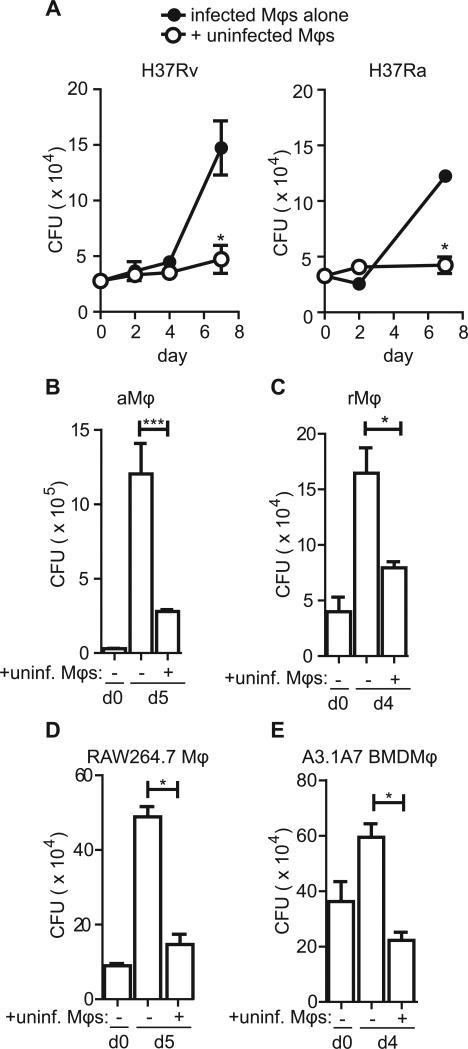
Figure 3. Uninfected macrophage co-culture limits bacterial growth.
(A,B) pMφs were infected with a 10:1 MOI of H37Rv (A) or H37Ra (B) for four hours before uninternalized bacteria were washed away and uninfected pMφs were added at a ratio of 2:1. Bacterial burden was assessed by CFU enumeration by plating serial dilutions of each condition in quadruplicate at the indicated timepoints. Data is representative of >20 experiments (A, H37Rv) and 3 experiments (B, H37Ra). (C) Alveolar Mφs (aMφs) were infected with an MOI of 10:1 H37Rv for 4 hours before the addition of uninfected aMφs (+unif. Mφs). CFU was enumerated as described on days 0 (d0) and day 5 (d5). Data is representative of 2 experiments. (D) Resident peritoneal Mφs (rMφs) were infected as described with H37Rv before the addition of uninfected rMφs and CFU enumeration on indicated days. Data is representative of 3 experiments. (E) RAW264.7 Mφs were infected with H37Rv as previously described prior to the addition of uninfected Mφs of the same type. CFU was determined. Data is representative of 2 experiments. (F) RAW264.7 Mφs were infected with H37Rv as previously described prior to the addition of uninfected Mφs of the same type. Data is representative of 2 experiments. Error bars ± SEM, p ≤ 0.05, Student's T-test.

