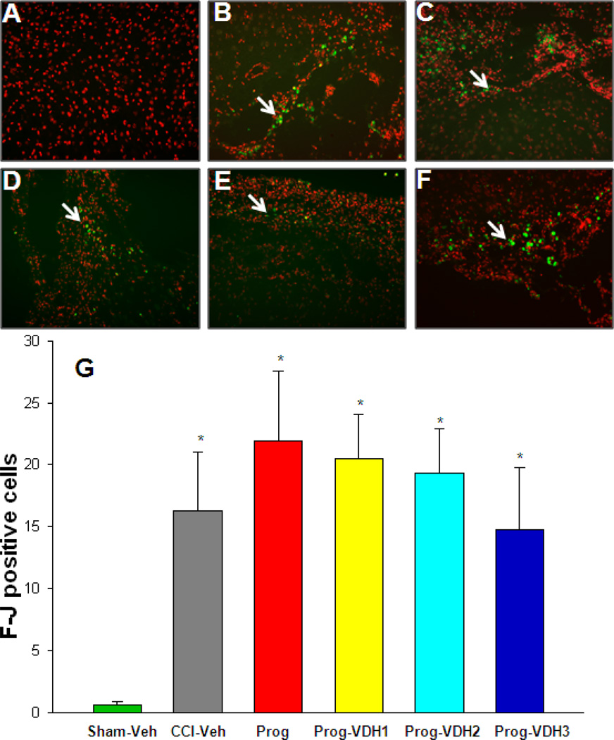
Figure 6. Degenerative neurons (Fluro-Jade staining).
Slides were stained with Floro-Jade C (F-Jc) antibody, and counterstained with propidium iodide (PI). The F-Jc positive staining appears as green (indicated by white arrows), and the PI as red. F-Jc positive cells significantly increased in all the CCI groups compared to Sham-Veh (*p<0.05). However, there were no significant differences between CCI-Veh, PROG and PROG+VDH groups. *Compared with Sham-Veh, p<0.05.

