Figure 1.
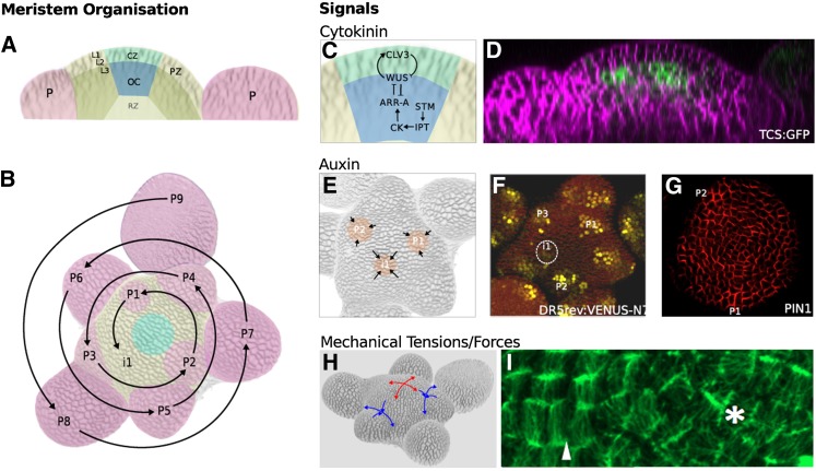
The Organized Structure of the Arabidopsis SAM Is Generated through a Combination of Hormonal and Mechanical Signals and the Cellular Responses to These Signals.
(A) The SAM consists of the central zone (CZ), containing the stem cells and the surrounding peripheral zone (PZ). Primordia (P), the progenitors of new lateral organs, are initiated in the PZ. The CZ is maintained by an underlying organizing center (OC). Below the OC is the rib zone (RZ), which is responsible for the elongation of the stem. In dicotyledonous species, the two outermost cell layers (L1 and L2) divide anticlinally and make up the so-called tunica layer. From the third cell layer (L3) inwards, cell divisions take place in nonuniform orientations, resulting in the less organized tissue structure known as the corpus. Organ initiation is largely controlled by auxin transport and distribution within the L1 of the tunica (see [E]), whereas maintenance of the CZ requires communication between the tunica and the corpus (see [C]).
(B) Primordia are spaced according to a regular pattern or phyllotaxis. In Arabidopsis, spacing follows a spiral phyllotaxy. P9 indicates the oldest primordium and P1 the youngest. Successive organs are separated by an angle of ∼137.5. The position at which the next primordium (i1) will be initiated can be predicted based on this rule.
(C) and (D) Cells in the OC express the transcription factor WUS, which promotes the expression of CLV3, a small peptide that moves into the surrounding tissue. In the central zone, CLV3 interacts with the receptor-like kinase CLAVATA1, inhibiting WUS expression and promoting stem cell fate. The hormone cytokinin (CK) is also required to establish and maintain the central zone. The transcription factor STM upregulates the expression of IPT genes that are rate limiting for cytokinin biosynthesis. Cytokinin activates A-type ARR transcriptional regulators via a phosphorelay system. A-type ARRs stimulate downstream cytokinin responses but also downregulate the expression of WUS. WUS inhibits the expression of A-type ARRs, creating a negative feedback loop that regulates size and position of the organizing center and, thus, of the central zone. Consistent with this role for cytokinin, the cytokinin response reporter Two-Component-Output-Sensor: Green Fluorescent Protein (TCS:GFP) (Müller and Sheen, 2008) (D) is detected at high levels in the organizing center, as previously shown by Yoshida et al. (2011).
(E) to (G) The positions of primordia are determined by auxin maxima (orange) that are created by self-organizing patterns of auxin transport (arrows) (E). The auxin response reporter DR5:3xVENUS-N7 is detected in primordia before outgrowth begins (see circled i1 in [F]). Directional movement of auxin is produced by the activity of PIN1 proteins, which transport auxin out of cells and are polarly localized. Immunolocalization of PIN1 in the L1 of the SAM shows that PIN1 proteins are oriented toward the auxin maxima (G).
(H) and (I) Mechanical principle directions of stresses experienced by the tissue varies across the SAM as a result of the geometry of the structure and as a result of local differences in growth rates. Stresses at the dome of the meristem are isotropic (red arrows), whereas in the fold between the outgrowing primordia and the SAM, the direction of stresses is highly anisotropic (blue arrows). Microtubules orient according to the principle direction of the mechanical stress that they experience. [I] shows a detail of a meristem expressing Microtubule Associated Protein4: Green Fluorescent Protein (as previously shown in Hamant et al., 2008). Note that microtubules at the meristem dome do not show a clear orientation (asterisk), but in organ boundaries a marked coordination of tubules is observed (arrowhead).
([D], F. Besnard and T. Vernoux, unpublished data, construct from Müller and Sheen [2008]; [G], reprinted from Barbier de Reuille et al. [2006], Figure 3F; [F] A. Jones, unpublished data, construct from Laskowski et al., 2008, [I], J. Traas, unpublished data, live imaging as described in Fernandez et al. [2010].)

