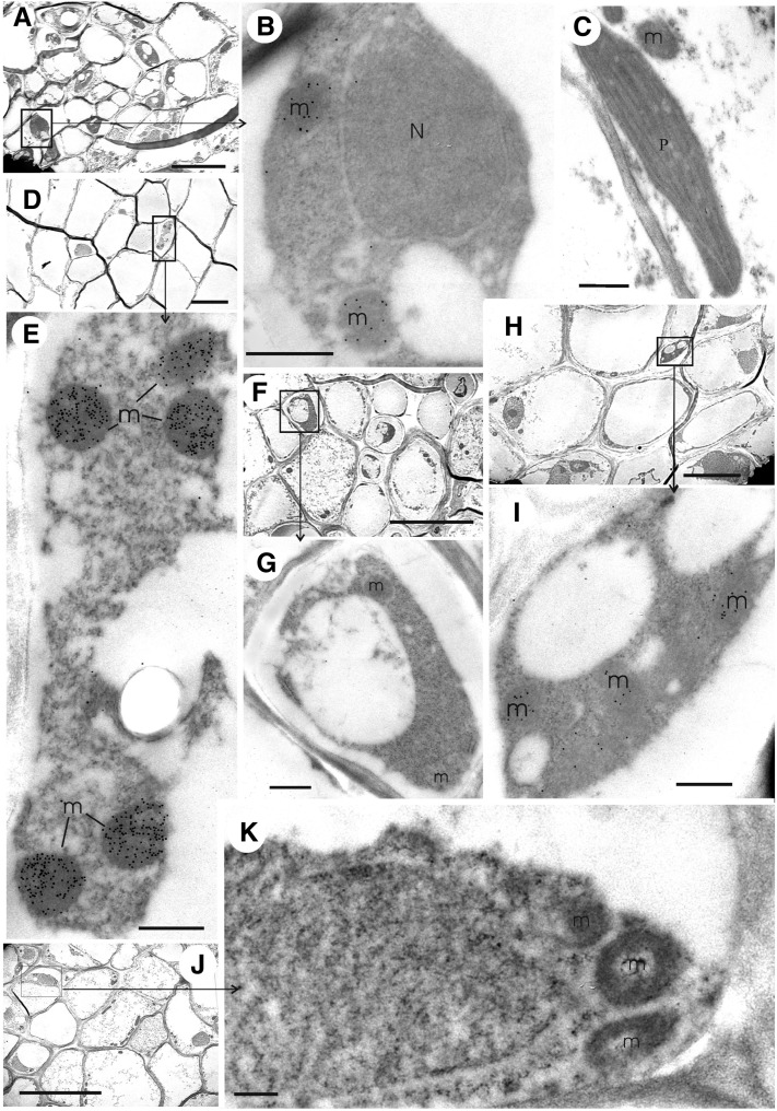
Figure 4.
Immunolocalization of NADH-GDH by Transmission Electron Microscopy in Roots and Leaves of the Wild Type and gdh1-2 and gdh1-2-3 Mutants.
Immunogold labeling experiments were performed on roots and leaves sections of the gdh1-2-3 mutant using the GDH antiserum raised against synthetic GDH polypeptides. m, mitochondrion; N, nucleus; P, plastid. Bars = 5 µm in (A), (D), (F), (H), and (J) and 0.5 µm in (B), (C), (E), (G), (I), and (K).
(A) Phloem tissues in wild-type leaves.
(B) Enlarged view of a companion cell in a leaf vascular bundle of the wild type (boxed area in [A]).
(C) Chloroplast and mitochondrion of wild-type parenchyma cell.
(D) Root phloem tissues in the wild type.
(E) Enlarged view of a companion cell in a root vascular bundle of the wild type (boxed area in [D]).
(F) Leaf phloem tissues in the gdh1-2 double mutant.
(G) Enlarged view of a companion cell in a leaf vascular bundle of the gdh1-2 mutant (boxed area in [F]).
(H) Root phloem tissues in the gdh1-2 double mutant.
(I) Enlarged view of a companion cell in a root vascular bundle of the gdh1-2 mutant (boxed area in [H]).
(J) Root phloem tissues in the gdh1-2-3 triple mutant.
(K) Enlarged view of a companion cell in a root vascular bundle of the gdh1-2-3 mutant (boxed area in [J]).

