Figure 3. .
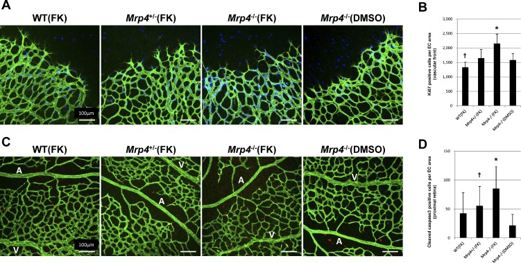
Intraperitoneal forskolin administration enhances both retinal EC proliferation and apoptosis in Mrp4-knockout mice. (A) Images of P6 retinas (at the vascular front) from forskolin- or DMSO-treated mice labeled for PECAM (green) and Ki67 (blue). (B) Quantification of the number of Ki67-positive cells per EC area. P value is < 0.01 in comparison between forskolin-treated Mrp4−/− mice and any other group (*). There is a significant difference between forskolin-treated WT mice and forskolin-treated Mrp4+/− mice or DMSO-treated Mrp4−/− mice (P < 0.01) (†). (C) Images of P6 retinas (in the proximal retina) from forskolin- or DMSO-treated mice labeled for PECAM (green) and cleaved caspase 3 (red). (D) Quantification of the number of cleaved caspase 3–positive cells per EC area. P value is <0.01 in comparison between forskolin-treated Mrp4−/− mice and forskolin-treated WT mice or DMSO-treated Mrp4−/− mice. There is a significant difference between forskolin-treated Mrp4+/− mice and DMSO-treated Mrp4−/− mice (P < 0.01) (†). A, artery; V, vein. The data are provided as the mean ± SD.

