
Figure 1.
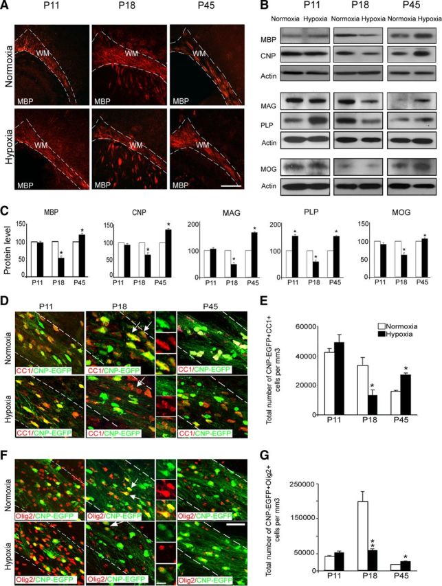
Hypoxia causes hypomyelination during white matter development. A, Confocal images of white matter from normoxic and hypoxic brains at P11, P18, and P45 immunostained with anti-MBP antibody. The dotted lines bound white matter. WM, White matter. Scale bar, 100 μm (n = 4 brains for each condition). B, Western blot analysis of white matter dissected from hypoxic and normoxic brains at P11, P18, and P45 demonstrates developmental expression of MBP, CNP, MAG, PLP, and MOG; actin was used as a loading control. C, Graphs represent transient changes in expressions of above proteins indicating a reduction in their level at P18, followed by an increase at P45 (n = 4 brains for each condition and for each antibody; *p < 0.05, t test). Confocal, tricolored images from normoxic and hypoxic white matter at P11, P18, and P45. CNP-EGFP+ cells are stained with anti-CC1 (D) and anti-Olig2 (F) antibodies. The dotted lines bound white matter. The insets magnify cells pointed by white arrows. Scale bar, 30 μm. Graphs represent total number of CNP-EGFP+ cells expressing CC1 (E) and Olig2 (G) in normoxic and hypoxic white matter at P11, P18, and P45. Hypoxia causes a significant reduction of CNP-EGFP+CC1+ and CNP-EGFP+Olig2+ cells at P18; however, a significant increase of oligodendrocytes is seen at P45. At P11, there was no difference in total number of oligodendrocytes in white matter after hypoxia (n = 4 brains for each condition, and for each antibody; *p < 0.05, **p < 0.02, t test). Error bars indicate SEM.