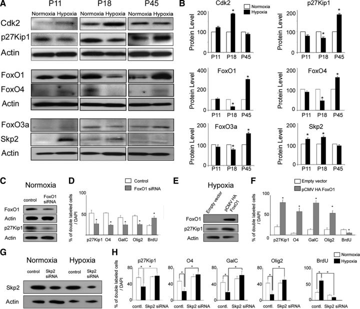
Figure 11.
FoxO1-dependent p27Kip1 expression in white matter OPCs after hypoxia. A, Western blot analysis of Cdk2, p27Kip1, FoxO1, FoxO4, FoxO3a, and Skp2 expression was performed in normoxic and hypoxic tissue at P11, P18, and P45. B, Graphs show expression levels of Cdk2, p27Kip1, FoxO1, FoxO4, FoxO3a, and Skp2 proteins quantified by Western blot. After hypoxia, Cdk2 and Skp2 levels were increased in white matter at P11 and P18. Conversely, p27Kip1, FoxO1, FoxO3a, and FoxO4 levels were decreased by hypoxia at P18, but were elevated at P45. Actin was used as a loading control (n = 4 for normoxia and hypoxia, respectively; *p < 0.05). C, D, FoxO1 loss of function in white matter cells in normoxic cultures. A reduction of 30–50% in FoxO1 and p27Kip1 levels was obtained after siRNA transfection. The graph shows the percentage of oligodendrocytes after FoxO1 siRNA and scrambled control transfection. In normoxic cultures, transfection with FoxO1 reduced the percentage of double-labeled EGFP+ cells expressing p27Kip1, O4, GalC, and Olig2, compared with cells transfected with scrambled control. However, the percentage of BrdU-proliferating cells was significantly elevated (n = 4 brains for each condition and for each antibody; *p < 0.05). E, F, FoxO1 gain of function in white matter cells after hypoxia. Western blot analysis demonstrates a fivefold increase in FoxO1 expression levels after transfection with pCMV5 HA FoxO1 plasmid, compared with empty vector transfection. In differentiation assays, FoxO1 overexpression caused an increase in the percentage of EGFP+ cells expressing p27Kip1, O4, GalC, and Olig2, and a reduction in proliferating cells, compared with cells transfected with empty vector (n = 4 brains for each condition and for each antibody; *p < 0.05). G, H, Skp2 loss of function in normoxic and hypoxic white matter cells. Western blot analysis demonstrates lower expression of Skp2 protein after Skp2 siRNA transfection in normoxic and hypoxic white matter cells, compared with transfection with scrambled control siRNA. In hypoxic cultures, transfection with Skp2 siRNA increased the percentage of double-labeled EGFP+ cells expressing p27Kip1, O4, GalC, and Olig2 oligodendrocytes, compared with normoxic cultures (n = 4 brains for each condition and for each antibody; *p < 0.05). Error bars indicate SEM.

