
Figure 13.
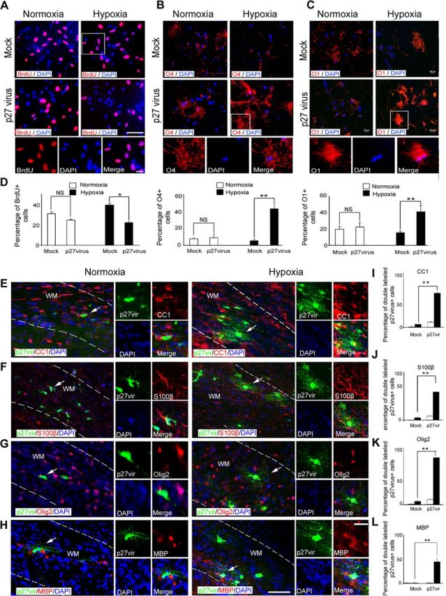
p27Kip1 overexpression restores oligodendrocyte differentiation after hypoxia. A, Cells from hypoxic and normoxic brains at P18 were transfected with p27Kip1 and mock retrovirus, and cultured 5 d in basal medium. For proliferation assays, cells were labeled with anti-BrdU antibody. The insets magnify cells in white boxes. Scale bar, 100 μm. For differential potential, cells were labeled with anti-O4 (B) and anti-O1 (C) antibodies. The insets magnify cells in white boxes. Scale bar, 100 μm. D, Graphs represent the percentage of BrdU+, O4+, and O1+ cells compared with all DAPI cells. p27Kip1 overexpression increased the percentage of O4+ and O1+ cells after hypoxia compared with mock-virus transfection. In normoxic cells, p27Kip1 overexpression did not change percentage of oligodendrocytes (n = 3 brains for each condition; *p < 0.05; **p < 0.02; NS, nonsignificant, t test). p27Kip1 enhanced differentiation potential of oligodendrocytes after hypoxia in vivo. In white matter, cells labeled with p27Kip1 retrovirus were colabeled with CC1 (E), S100β (F), Olig2 (G), and MBP (H) markers after normoxia and hypoxia. The insets magnify cells pointed by white arrows. WM, White matter. Scale bar, 50 μm. Graphs represent percentage of double-labeled p27virus+ cells stained with CC1 (I), S100β (J), Olig2 (K), and MBP (L) markers. After hypoxia, cells transfected with p27Kip1 retrovirus expressed Olig2, CC1, and S100β. In normoxia, p27Kip1 did not significantly change the number of mature oligodendrocytes compared with mock virus controls (n = 4 brains for each condition, and for each antibody; *p < 0.05, two-way ANOVA). Error bars indicate SEM.