
Figure 2.
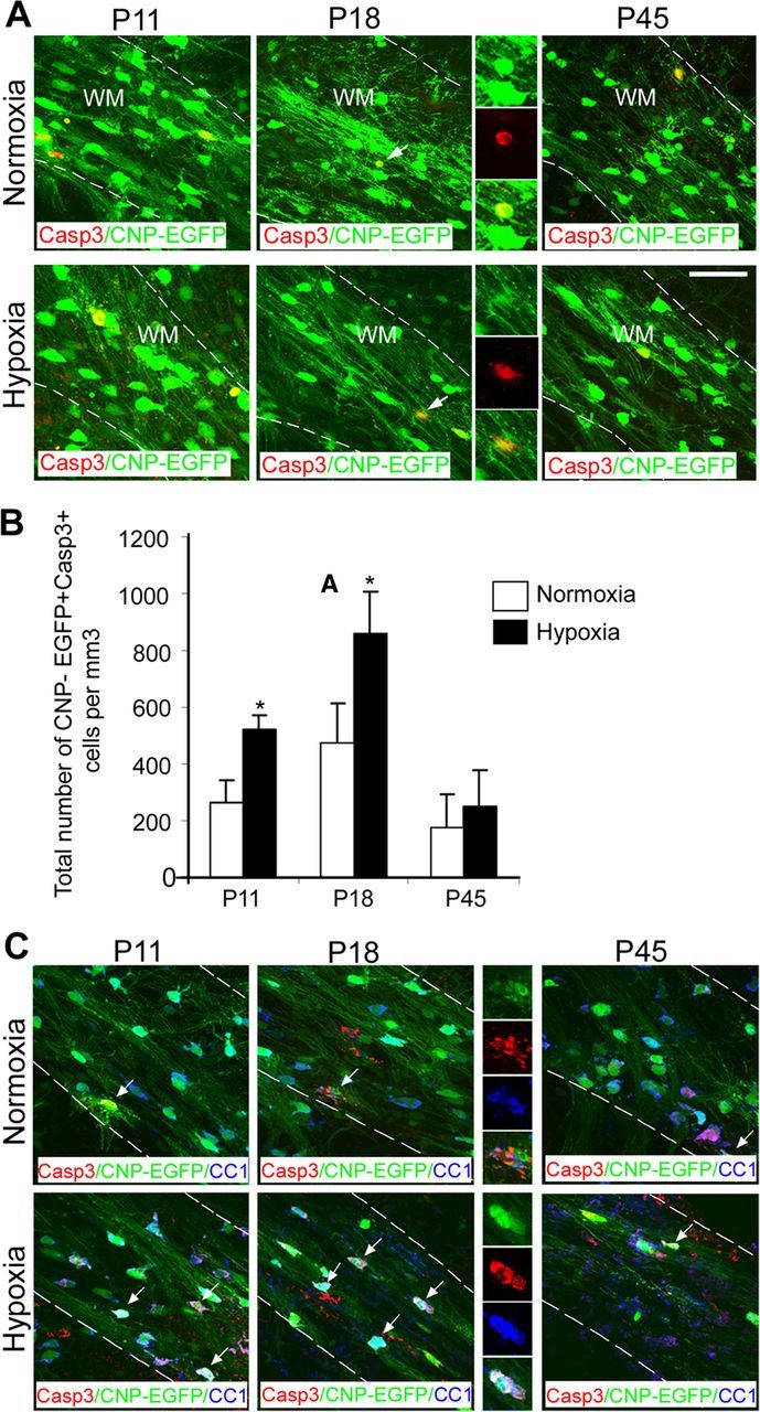
Hypoxia increases apoptosis of oligodendrocytes. A, Confocal images from normoxic and hypoxic white matter. CNP-EGFP+ cells were doubled-stained with anti-Caspase 3 antibody. The dotted lines bound white matter. The insets magnify cells pointed by white arrows. WM, White matter. Scale bar, 50 μm (n = 4 brains for each condition). B, Increased apoptosis of CNP-EGFP+ cells in white matter was observed at P11 and P18 (n = 4 brains for each condition; *p < 0.05, t test). C, Confocal images from normoxic and hypoxic white matter. CNP-EGFP+ cells were triple-stained with anti-Caspase 3 and anti-CC1 antibodies. The dotted lines bound white matter. The insets magnify cells pointed by white arrows. WM, White matter. Scale bar, 50 μm. Error bars indicate SEM.