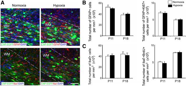
Figure 6.
Hypoxia does not affect astrocyte and microglia in white matter. A, Confocal images from normoxic and hypoxic white matter at P18 demonstrating astrocytes labeled with GFAP and microglia labeled with Iba1. The dotted lines bound white matter. WM, White matter. Scale bar, 100 μm. The graphs present total number of GFAP+ and GFAP+Ki67+ (B), Iba1 and Iba1+BrdU+ (C) at P11 and P18. No difference in total number of astrocytes and microglial cells, and their proliferation were detected in white matter immediately and 1 week after hypoxia (n = 4 brains for each condition and for each antibody). Error bars indicate SEM.

