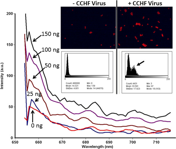
Figure 4.

Results of the two-fold serial dilution experiment using formalin-fixed IbAr 10200 strain of CCHF. Excitation was at 645 nm with 5 nm slits and emissions were scanned from 655 to 720 nm with a PMT setting of 900 V. The inset shows fluorescence microscopic images captured from the zero control (− CCHF virus) and the 150 ng of inactivated virus (+ CCHF virus) samples scraped from the inside of cuvettes and placed on microscope slides. NIH Image J image analysis software was used to verify that the red TYE 665 fluorescence intensity increased after capture of 150 ng of CCHF virus as illustrated by the histograms in the inset (highlighted by the arrow).
