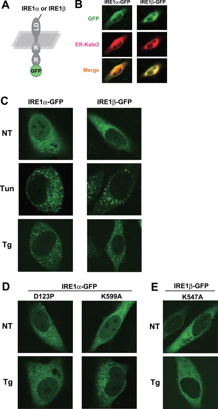
Figure 1. Clustering of IRE1α and IRE1β upon ER stress.
(A) Schematic of IRE1-GFP imaging construct. Luminal domain (“LD”), kinase domain (“K”), RNase domain (“R”) of human IRE1s, and fused monomer-GFP are indicated. (B) ER-localization of GFP-fused IRE1s. Fluorescent images were collected from GFP-fused IRE1s and ER-mKate2 construct – transfected HeLa cells. (C) Clustering of IRE1α and IRE1β upon ER stress. GFP-fused IRE1s were transfected into HeLa cells and treated with or without tunicamycin (2.5 µg/ml for 2 h) or thapsigargin (1 µM for 2 h), after which fluorescent images were collected. (D, E) Clustering defect in mutant IRE1s. GFP-fused IRE1α (D123P or K599A) (D) or GFP-fused IRE1β (K547A) (E) were transfected into HeLa cells and treated with or without thapsigargin (1 µM for 2 h), after which fluorescent images were collected.

