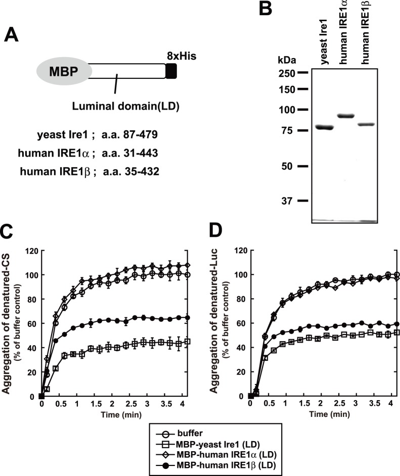
Figure 3. Anti-aggregation activity of IRE1β.
(A) Schematic of recombinant fragments used in the anti-aggregation assay. (B) SDS-PAGE of recombinant fragments used in the anti-aggregation assay. Bacterially expressed fragments were purified by Ni-NTA, run on 8% SDS-PAGE gels, and stained with Coomassie blue. (C, D) Anti-aggregation assay with the recombinant fragments. At time 0, citrate synthase (C), luciferase (D) in guanidine HCl-denaturing buffer were diluted into assay buffer, with or without each recombinant fragment. Turbidity of the sample mixtures was monitored by measuring absorbance at 320 nm and normalized against the maximum value of the buffer sample. The average and SEM from three reactions are shown.

