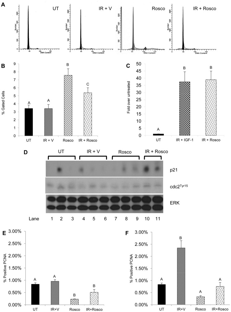
Figure 1. Increased cell cycle arrest in irradiated parotid glands pretreated with Roscovitine.
The head and neck region of female FVB mice was treated with a 5Gy dose of radiation with or without 100 mg/kg Roscovitine pretreatment. Parotid glands were removed 6 hours after radiation treatment. A) Representative flow cytometry histograms from untreated (UT), irradiated with vehicle pre-treatment (IR+V), Roscovitine alone (Rosco), and Roscovitine prior to irradiation (IR+Rosco). B) Tissues were dispersed, stained with propidium iodide, and analyzed by flow cytometry. Graphical representation of the mean percentage of cells gated in G2/M with SEM from ≥9 mice per treatment. C) RNA was isolated from parotid glands, treated as stated above and real-time RT-PCR was run with primers for p21 amplification. Results were calculated using the 2−ΔΔCt, normalized to untreated and displayed as mean with SEM≥4 mice per treatment. D) Parotid tissues were treated as stated above and collected 6 hours following radiation for immunoblotting and prepared as described in the materials and methods, and membranes were probed for p21 (top panel) and cdc2Tyr15 (middle panel), with total ERK (bottom panel) as a loading control. Tissues were collected at 24 hours (E) and 48 hours (F) from irradiated mice pretreated with Roscovitine, embedded in paraffin, and stained for PCNA as described in the materials and methods. Results are graphed as the number of PCNA-positive acinar cells as a percentage of total counted acinar cells. The data are shown as the mean with SEM≥3 mice per treatment group. Treatment groups with the same letters are not statistically different from each other.

