Figure 4.
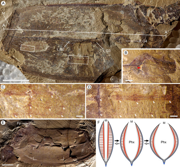
Gill anatomy and body-wall musculature of Vetulicola rectangulata. (A) Laterally compressed specimen ELI-SJ0605A, with preserved muscular fibers beneath the plate-like unit; note also the tightness of the lateral groove. (B) Internal view of right gill slit 4 in (A); note the internal opening (red arrowhead) surrounded by striations. (C,D) Enlargement of boxed areas in (A) at left and right side, respectively, showing fibrous structures interpreted as longitudinal muscles, defined by diagenetic iron minerals; note their location beneath the plates and arrangement as continuous bundles (approximately six fibers per bundle) extending almost the entire length of the anterior section; note also the equidistant expansions (arrowheads), interpreted here as corresponding to a column of horizontal muscles. (E) Specimen ELI-0000306, showing inferred muscular imprints represented by longitudinal structures in the anterior section (notably the focused area). (F) Reconstructed horizontal section (membrane in blue, plates in grey, musculature in red), equivalent to section A-A' in (A), demonstrating inferred dynamics of pharyngeal dilation, governed by the collective action of the longitudinal muscle fibers and their horizontal derivatives. Abbreviations: Co, cowl-like structure; Fp, fin-like process; LG, left gill slit; M, mouth; Phx, pharynx; RG, right gill slit; T, tail; Wf, water flow. Scale bars: 1 cm in (A), (E); 1 mm in (B) to (D).

