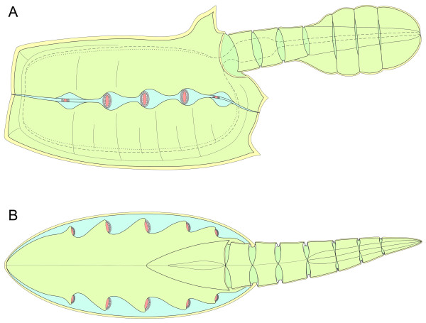
Figure 7.
Schematic reconstruction of Vetulicola rectangulata. (A) Lateral view. Lappets of left gill 2 to 4 removed to reveal gill openings and surrounding features; pharynx and alimentary canal denoted by dashed line; dorsal and ventral food grooves denoted by dotted lines; gill slits colored in pink. (B) Dorsal view. Semidiagrammatical image depicting the internal arrangement of the gills. The diagram is principally to show the overall arrangement of the gills with respect to the rest of the body. Gill morphology is more complex than the diagramatic depiction provided here, and is illustrated in detail in other Figures.

