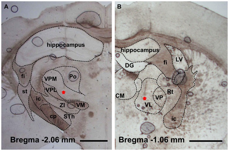
Figure 1. Verification of electrodes location.
Electrode placements in ventral posteromedial (A) and ventral lateral (B) nuclei were histologically verified with light microscopy. Arrow point indicates the tip of electrode. fi, fimbria of the hippocampus; st, stria terminalis; ic, internal capsule; cp, cerebral peduncle (basal part); VPM, ventral posteromedial nucleus; VPL, central posterolateral nucleus; ZI, zona incerta; STh, subthalamic nucleus; Po, posterior thalamic nuclear group; VM, ventromedial thalamic nucleus; DG, dentate gyrus; CM, central medial nucleus; VL, ventral lateral nucleus; VP, ventral posteromedial and posterolateral nuclei; Rt, reticular thalamic nucleus; LV, lateral ventricle. Scale bar is 1 mm.

