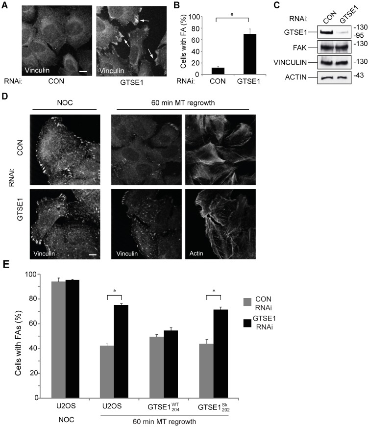
Figure 5. GTSE1 modulates focal adhesion disassembly in an EB1-dependent manner.
(A) Immunofluorescence of U2OS cells transfected with control (CON) or GTSE1 siRNA for 24 h followed by serum starvation for 48 h, stained for vinculin. Focal adhesions persist in cells depleted of GTSE1. Scale bar represents 10 microns. (B) Quantification of focal adhesion (FA) disassembly from experiments from (A). The percentage of cells containing 10 or more focal adhesions after serum starvation-induced disassembly was determined (n = >50 cells per experiment, 3 experiments for each condition). * indicates p<0.05 as determined by a Student’s t test. (C) Western blot of U2OS cells transfected with control (CON) or GTSE1 siRNA and serum-starved for 48 h, blotted with anti-GTSE1, anti-FAK, anti-vinculin, and anti-actin. (D) Immunofluorescence of U2OS cells transfected with control (CON) or GTSE1 siRNA for 36 hours. Cells were imaged after treatment with nocodazole for 4 hours, and 60 minutes following washout of nocodazole to allow microtubule regrowth. Cells are stained for vinculin and actin. GTSE1-depleted cells contain more focal adhesions that wild-type following microtubule regrowth. Scale bar represents 10 microns. (E) Quantification of focal adhesion disassembly in U2OS cells, following the assay described in (D). Cells stably expressing RNAi-resistant wild-type GTSE1-GFP (GTSE1WT 204) or GTSE1-GFP mutated at the SKIP motifs (GTSE1Sk 202) were additionally assayed. Quantification was performed as described in (B). Cells containing only mutant GTSE1 unable to interact with EB1 or track growing microtubule ends are deficient for microtubule-dependent focal adhesion disassembly.

