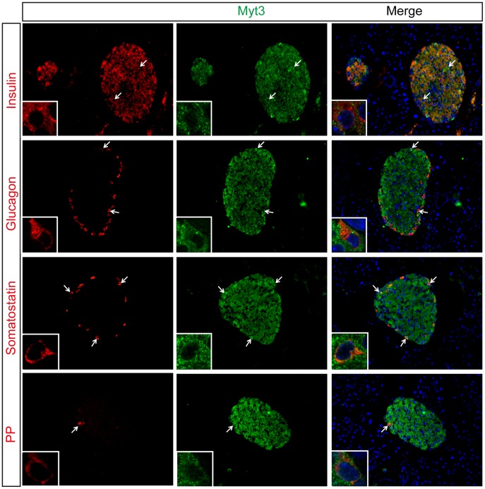
Figure 4. Myt3 co-localises with endocrine cell markers in adult pancreas.
Saggital sections of adult pancreata were analysed for expression of Insulin, Glucagon, Somatostatin and Pancreatic Polypeptide, as indicated (red), and Myt3 (green). Nuclei were stained with Hoechst (blue). Arrows indicate co-localisation of Myt3 with indicated endocrine markers. High magnification confocal images of individual cells showing co-localization of Myt3 with Insulin, Glucagon, Somatostatin and Pancreatic Polypeptide, representative cells are shown (Inset), note that Myt3 staining is predominately cytoplasmic but can also be found within the nucleus.

