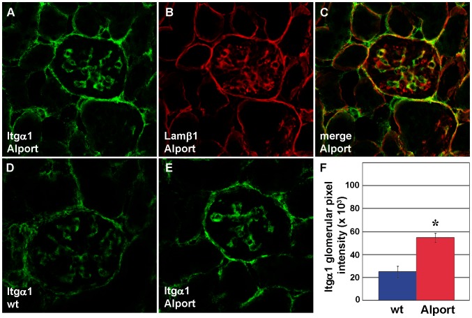
Figure 4. Integrin α1 protein is upregulated in the mesangium of Alport glomeruli.
A–C: Fresh frozen kidney sections from 4 week old Alport mice were labeled with a combination of hamster anti-integrin α1 and rat anti-laminin β1 IgGs, followed by the appropriate species-specific Alexa Fluor secondary antibody. Anti-integrin α1 labeling (A, Itgα1) is restricted to the mesangial layer, marked by anti-laminin β1 staining (B, Lamβ1), and overlap of staining is shown in C (merge). D–F: Representative fluorescence micrographs are shown of anti-integrin α1 labeling of wild-type (D, wt), or Alport (E) mouse glomeruli. Glomerular fluorescence intensities were averaged for n = 3 mice of each genotype, wildtype (wt, blue) or Alport (red), and integrin α1 signals were significantly greater in Alport. * p = 0.01.

