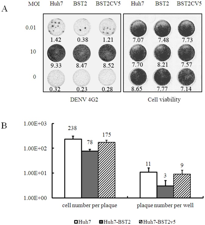
Figure 4. BST2 inhibits DENV spread via cell-to-cell transmission.
The cells were infected with DENV at a MOI of 0.01 or 10 for 1 h and culture media were replaced with media containing 0.5% methocellulose to prevent cell-free virus infection and cultured for 2 days. (A) Representative DENV-infected cell foci from cultures of the three cell lines. The infected cell foci and cell viability were revealed by In-Cell Western assay by using of antibody against DENV E protein and Sapphire 700 staining, respectively. The indicated gray values of the dots were quantified by using of an Odyssey Infrared Imaging System (LI-COR Biotechnology). (B) The average infectious foci number per well in 24-well plate and the average DENV-infected cell number per focus from 100 foci were plotted. (C) The intracellular DENV RNA was determined for the cells infected with DENV at MOI of 10 by qRT-PCR assay. The values were presented as percentage of values from the Huh7-BST2 and Huh7-BST2CV5 cells compared with that from parent Huh7 cells. The experiment was performed in 3 replicates to generate statistically sufficient data. p values were calculated using Student's t test.

