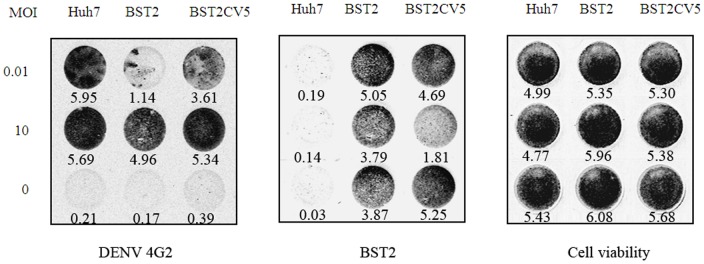
Figure 5. In-cell western analysis for DENV infection in Huh7-BST2 and Huh7-BST2CV5 cells.
Cells were infected with DENV at indicated MOI and cultured for 2 days with complete medium. Cells were fixed and double-staining of DENV 4G2 protein and BST2 were revealed by In-Cell western assay. The indicated gray values of the dots were quantified by using of an Odyssey Infrared Imaging System (LI-COR Biotechnology). The values represent average from 3 independent experiments.

