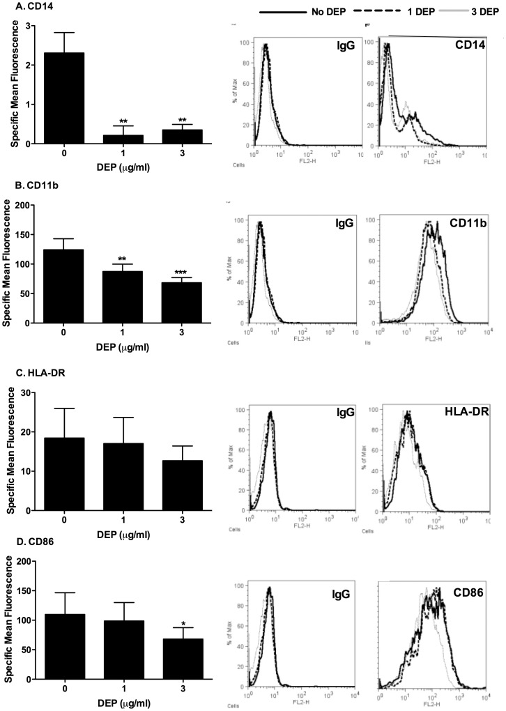
Figure 8. MDMs differentiated in the presence of DEP showed reduced CD14, CD11b and CD86 surface marker expression.
MDMs from healthy volunteers were differentiated from purified monocytes in the presence or absence of varied concentrations of DEP as described in the methods. At day 14 MDMs were harvested and non specific binding was blocked by incubating at room temperature for 10 minutes with 1∶50 IgG from murine serum reagent grade in FACS buffer. Cells were then incubated with 1∶10 PE conjugated mouse anti-human CD14, CD11b, HLA-DR, TLR2, TLR4, CD80 or CD86 or the respective isotype controls. PE fluorescence was measured on the FL-2 channel of a FACSCalibur flow cytometer. Data are presented as a mean±SEM of n = 4 performed on different donors and representative histogram flow analysis plots from one donor. Significant differences between MDMs and DEP-MDMs cell surface expression are denoted by *p<0.05 and **p<0.01 as measured by one way ANOVA and Dunnett’s post test.

